Causes of cardiomyopathy

Causes of cardiomyopathy


Pregnancy-related cardiomyopathy

What causes peripartum cardiomyopathy?
The cause and the underlying disease mechanisms of peripartum cardiomyopathy is unknown, and many theories have been proposed. Nutritional disorders, autoimmune disease, inflammatory disease, viruses, blood cells from the fetus that enters the mother's blood as well as other theories have not led to any conclusion about the cause or progression of disease.

We are also uncertain about what might predispose to this disease. It is suggested that the following factors may increase the risk: high maternal age, number of previous births, African origin, pre-eclampsia, high blood pressure, use of rihemmende agent and twins.
Causes of cardiomyopathy.jpg

Despite this, we know about. 1/3 of the cases occur among young nulliparous.

Pregnancy related cardiomyopathy

Pregnancy related cardiomyopathy


What is pregnancy-related cardiomyopathy?

Pregnancy-related cardiomyopathy (peripartum cardiomyopathy) is a condition of unknown cause in which early signs of heart failure and symptoms of heart failure occur between the last month of pregnancy and the first five months after birth. However, there are reports of similar disease pictures earlier in pregnancy, cases may be due to this condition.

Cardiomyopathy means that the disease attacks the muscles of the heart. Peripartum means surrounding the birth. The diagnosis of peripartum cardiomyopathy is made by excluding other causes of cardiomyopathy.
Pregnancy related cardiomyopathy

A heart that fails in this condition will cause the heart expands - dilated - is greater. It increases the risk of blood clots can form in the heart. Such blood clots can break loose, float in the blood and clog blood vessels elsewhere in the body.

Peripartum cardiomyopathy is a relatively rare disease. There are no reliable prevalence data, and the numbers that exist, differ widely. In the U.S., it is suggested that there are approx. 1 case per 4000 births.

Treatment for hypertrophy

Treatment for hypertrophy


Preventive treatment

Young people with known hypertrophic cardiomyopathy should be advised not to expose themselves to excessive exertion, even if their condition is asymptomatic.

They should therefore, not participate in competitive sports with high standards of fitness. Most, however, participate in recreational activities that cause only mild to moderate physical strain.

How are long-term prospects?

Many patients may be asymptomatic throughout life and have a normal life. Among those who will develop these symptoms usually slowly. Over time, it can develop heart failure or angina pectoris.
Treatment for hypertrophy

Sudden death can occur, but modern treatment with the defibrillator and cardiac surgery aims to reduce the risk of such events.

Treatment for hypertrophic cardiomyopathy

Treatment for hypertrophic cardiomyopathy


What is the treatment?

The goal of treatment is to reduce symptoms and possibly prevent complications, particularly sudden death.

People without symptoms, and where there is no information on family cases of unexpected death at a young age, should not be treated medically, but is followed by a heart specialist.

Drug therapy is the primary treatment if the patient has symptoms. In complicated cases where medicines are working poor, both pacemaker therapy and surgical treatment are appropriate.
Treatment for hypertrophic cardiomyopathy

In some cases, a defibrillator implanted under the skin and in extreme cases, heart transplantation may be required.

Hypertrophic cardiomyopathy diagnosis

Hypertrophic cardiomyopathy diagnosis


How is the diagnosis?
Medical history and any ECG changes suggestive of cardiac disease. In the further investigation is echocardiography, ultrasound of the heart, the main survey. Here, the doctor can see the thickening of the heart wall, and any abnormal pressure differences in the heart can be measured. When your doctor does not find evidence of another disease that could explain the enlarged heart, we can conclude that there is hypertrophic cardiomyopathy.

Other investigations that may do are exercise-test and 24-hour heart rate monitoring. Sometimes also made ​​MRI of the heart. Genetic tests are of little use.

It is recommended that first-degree relatives (parents / siblings / children) examined the diagnosis in a person. Although family members do not have symptoms or signs of disease, it may still be changes in the heart.
Hypertrophic cardiomyopathy diagnosis

It is usual to follow the relatives of controls (ECG and echocardiography) every other year in young adulthood and every five years in adulthood.

Signs and symptoms of hypertrophic cardiomyopathy

Signs and symptoms of hypertrophic cardiomyopathy


Symptoms and signs
Patients with hypertrophic heart disease, for many years without symptoms of heart disease. If symptoms recur, the most common breathing difficulty and chest pain on exertion ( angina ).

Some brand's palpitations or anxious heart. Some shy of dizziness and fainting, often triggered by exertion.

Examination by a doctor may not be very wrong to find in the early stages. However, there may be a murmur over the heart.

ECG , however, usually not normal. It often shows signs of altered flow distribution in the heart (conduction disorder) and various degrees of thickened muscle of the left ventricle.
Signs and symptoms of hypertrophic cardiomyopathy

Sometimes one can find atrial fibrillation or other arrhythmias.

Cause of hypertrophic cardiomyopathy

Cause of hypertrophic cardiomyopathy


What is the cause of hypertrophic cardiomyopathy?

It Must have a family history of known cases of heart disease and sudden cardiac death. The condition is hereditary and is caused by mutation (error) in one or more genes. Gene's defect leads in turn to the muscle cells of the heart grows too much.

The changes in the heart can vary from person to person. Many small changes and have no complaints. Typically, the wall of the left ventricle (ventricle), which is thickened (hypertrophic). At 20-30% will be the end of the left ventricle cramped, making it difficult for the heart to pump blood out into the wide circulation. Furthermore, anterior chamber (atria) may eventually be enlarged.
Cause of hypertrophic cardiomyopathy

The changes in heart muscles and the atria predisposing to arrhythmias in the heart. In some cases, these arrhythmias may be severe and lead to cardiac arrest.

What is hypertrophic cardiomyopathy

What is hypertrophic cardiomyopathy


Enlarged heart - hypertrophic cardiomyopathy

Cardiomyopathy means disease of the heart muscle. Hypertrophy means enlargement. Hypertrophic cardiomyopathy is a disease where the heart muscle is enlarged, and were in some cases lead to symptoms due to impaired cardiac function.

Typically, an enlarged heart due to underlying disease such as congenital valvular abnormalities or high blood pressure. However, the condition hypertrophic cardiomyopathy is no such underlying disease. The disease is hereditary and is due to changes in genes (mutations) that can be transmitted to new generations.
What is hypertrophic cardiomyopathy

Hypertrophic cardiomyopathy occurs in approximately. 1 of 500-1000 adults. The disease is present from childhood, but rarely produces symptoms until puberty. Often it is discovered wounded in youth or adulthood. The condition is the most frequent cause of sudden cardiac death among young people and athletes.

Hyperlipidemia treatment guidelines

How is hyperlipidemia?


Treatment goals
If it is necessary to treat, apply the following treatment goals for fats in the blood:

  • Total cholesterol less than 5.0 (mmol / L)
  • Relationship between the values ​​for total cholesterol and HDL cholesterol (good cholesterol) should be less than 4
  • LDL cholesterol should be lower than 3.0 (mmol / L)
  • Triglycerides should be less than 2.0 (mmol / L)
  • HDL cholesterol should be higher than 1.0 (mmol / L)

General treatment
Total risks for heart disease are essential for the gain of fat-lowering treatment. A slightly elevated cholesterol is isolation tungveiende no reason for treatment; it is the sum of risk factors is basic. That is, the more of the factor's hyperlipidemia, hypertension, diabetes, genetic predisposition, smoking, obesity, lack of exercise that is, the more important it is to manage to avoid the development of cardiovascular disease.

All treatments start with a change of diet. The next step in treatment is the use of fat-lowering drugs, called statins. In some cases, it may also be appropriate to use triglyceride funds.

Dieting recommended for obesity. Smoking is a major risk factor for vascular disease of the heart and smoking cessation is therefore, important. Regular exercise improves the composition of fats in the blood. It is often necessary to get a permanent weight loss. The goal is at least 30 minutes of exercise 2-3 times a week.
Hyperlipidemia treatment guidelines

The isolated elevated triglycerides recommended low alcohol intake, caution with coffee, weight loss in overweight people.

What if you have elevated cholesterol, but otherwise no other risk factors?
  • When cholesterol levels between 6.5 to 7.9 and LDL-cholesterol of 5.0, or a relationship between LDL and HDL over 5, the non-drug therapy
  • Cholesterol of 8.0 is treated first with drugs.
  • If LDL-cholesterol continues to be above 5.0, or the relationship between LDL and HDL continues to be above 5 at follow-up, drug therapy should be considered.
  • Women are not treated medically until after they have ceased to menstruate.

Self-Treatment
The restructuring of lifestyle should continue for at least six months before taking the drug therapy. The exceptions are patients with heart disease, which often starts with drug treatment and lifestyle change simultaneously.

Dietary advice
The amount of saturated fats in food is reduced to less than 10% of the total energy (calorie intake). The main sources of saturated fat in the diet are meat, meat products (15-20%), margarine and milk products (40%). Limit your total fat intake to 30% of energi-/kaloriinntaket Cover 50% of energy intake from carbohydrates - cereals, fruits and vegetables. Increase your intake of fatty fish, soft margarines and olive oil. Have a moderate alcohol consumption. A intermediate consumption of nuts, 50-100 g daily, can reduce cholesterol levels by approx. 0.3 mmol / l.

Pharmacotherapy
Cholesterol-lowering medications given to patients without known heart disease if they are at high risk of heart attack.

In patients with known cardiac disease or symptomatic disease that confined Halske, stroke, or poor blood circulation, offered cholesterol-lowering drugs to patients with cholesterol levels below 5.5. The decision on the treatment a physician and patient assess the benefits, and the side effects and disadvantages of treatment can have. Treatment is often up to 65-75 years of age. The benefit in the elderly is under discussion.

Familial hypercholesterolemia

It is reasonable to believe that adult family members with LDL-cholesterol above 5.5 have familial hypercholesterolemia. Treatment Start from 18 years of age or earlier are implemented at high risk for vascular disease of the heart.

Isolated elevations in triglycerides

Consideration should be given medical treatment when the condition in the family, when the condition is combined with early heart disease and when triglycerides are over 10, because. Increased risk of inflammation of the pancreas.

Hyperlipidemia diagnosis

Hyperlipidemia diagnosis


How hyperlipidemia diagnosed?
It is important to clarify whether there is an accumulation of hyperlipidemia in the family. Are there relatives with elevated cholesterol? Are there cases of early heart disease (before age 60 years) in the family? User patient medication that may affect the lipid profile? If there are chronic diseases that may contribute to hyperlipidemia? Since hyperlipidemia is included as one of the several risk factors for heart disease, your doctor will also determine smoking habits, eating habits and alcohol intake.

The study sees the doctor for fat deposits in the eye area and around tendons. In order to assess the patient's overall risk of developing heart disease, your doctor will measure blood pressure, weight, listen to the throat, kidneys and groin arteries; feel for the pulse in the groin and lower legs and work out the relationship between livmål and hip measurement. This should be below 1.0 in males and below 0.9 in women.
Hyperlipidemia diagnosis

Measurement of fats in the blood is essential for diagnosis. This means the measurement of total cholesterol, HDL-cholesterol and triglycerides (fasting). The value for the dangerous part of the cholesterol (LDL cholesterol) can be calculated if one knows the values ​​mentioned above. Measurement of blood glucose is done to see if it could be diabetes. The cholesterol of 8 and isolated occurring elevations in triglycerides are taken in addition to glucose metabolism also hormones, liver test, kidney test and urine test strip examined for the presence of proteins (kidney damage). In many cases, your doctor will as well take a heart (ECG) to check for signs of damage of the heart.

How are long-term prospects?
Fat values ​​vary with age and gender. Fats appear to increase by 2-3 mmol / L from 20-30 years of age to 60-70 years of age. Long-term prospects depend on cholesterol levels, but primarily of the total risk of vascular disease of the heart. The complication of hyperlipidemia is heart disease.

Hyperlipidemia risk factors

Hyperlipidemia risk factors


Metabolic Syndrome
This is a condition with normal or slightly overhead total cholesterol. HDL cholesterol is reduced, and triglycerides are elevated. At the same time, these patients have elevated blood pressure and decreased tolerance for sugar, which means that many develop diabetes ( diabetes ). Another typical feature of these patients is obese around the abdomen.

Other issues
A number of diseases and medications can also contribute to the development of hyperlipidemia. It can apply to diabetes mellitus (diabetes), disorders of the thyroid gland ( hypothyroidism), kidney and liver disease, alcohol consumption, use of certain water pills; beta blockers may be unfavorable (heart medicine), cortisone drugs, and estrogens (female sex hormone).

What causes hyperlipidemia

What causes hyperlipidemia


Primary hyperlipidemia
The condition is due to inheritance of multiple genes that predispose to high cholesterol. In addition, the condition is often the lifestyle related. Cholesterol levels are moderately elevated and are usually around 6-8 mmol / L. The pattern of inheritance is such that hyperlipidemia occur scattered in the family. This means that not everyone in the family inherits the facility for the disease.

Familial hypercholesterolemia
This is called a dominant hereditary disease. This means that it is likely to inherit the condition, and the risk can be calculated as follows:
  • If one parent has a healthy and a diseased gene (called heterozygous) and the other parent is healthy, there are a 50% chance that a child inherits the disease.
  • If one parent has two diseased genes (called homozygous) and the other parent is healthy, all kids get sick.
  • If both parents have a diseased gene, there are a 75% chance that a child inherits the disease.
  • If both parents have two diseased genes, all kids get sick (and all will be homozygous)
What causes hyperlipidemia

The disease is characterized by the formation of grease balls in the eye area and along the tendons. The disease affects 1 in 400, but accounts for 5% of all cases of myocardial infarction. These individuals are therefore, at high risk for vascular disease of the heart. In the heterozygote, the complete cholesterol often be between 8 and 12 mmol / L. In homozygous, the total cholesterol eventually be above 20 mmol / L.

Familial combined hyperlipidemia.
The condition found in 1 of 50-100. It provides the same risk of disease in the heart as familial hypercholesterolemia. Total cholesterol is usually of 7-9 mmol / L, and triglycerides are above two mmol / L.

What is hyperlipidemia

What is hyperlipidemia?
Hyperlipidemia has high levels of fats in the blood. The term used for the increase of lipid cholesterol and triglycerides. Cholesterol can be measured as total cholesterol, HDL-cholesterol and LDL-cholesterol. HDL cholesterol is often termed "good cholesterol" because high HDL-cholesterol had been shown to protect against heart disease. Conversely, low HDL and high LDL cholesterol have an adverse effect on the heart.

Total Cholesterol
Limits of cholesterol (the concentration in blood is measured in mmol / L):

Less than 5, the desired level
5.0 to 6.4 implies easily elevated cholesterol
6.5 to 7.9 means moderately elevated cholesterol
Higher than 8.0 means expressed elevated cholesterol
Triglycerides
Triglyceride level higher than 2.3 mmol / L is considered elevation.
What is hyperlipidemia

Lipids as a risk factor
There is a clear association between cholesterol levels and risk of developing diseases of the heart. Elevated levels of triglycerides seem to be an independent risk factor for developing vascular disease of the heart, particularly in women.

Occurrence
In Norway, about 200,000 people a familial hypercholesterolemia in which some of the family has elevated cholesterol and where an inheritance pattern is related to several genes. Closely, 50,000 people have been familial combined hyperlipidemia in which both cholesterol and triglycerides are upraised. Approximately, 10,000 people have elevated cholesterol caused by a defect in a specific genetic material.

Pulmonary embolism prognosis

Pulmonary embolism prognosis


How are the course and prognosis?
In general, because the disease decided. Any worsening of cor pulmonale depends upon both on how early treatment starts and how effective this is. Complications are right-sided heart failure and right-sided heart attack.

Good treatment of grunnsykom well as the reduction of oxygen is important to avoid further forverrring of secondary pulmonary hypertension. The prognosis depends on the underlying disease. For example, patients with COPD and moderate respiratory prevent a 3-year mortality risk of 50% right-sided heart failure after occurred nine.

Pulmonary embolism prognosis
In patients who live many years with their basic disease, it may eventually develop cor pulmonale and right-sided heart failure If the patient develops right-sided heart failure, this could be treated on equal terms with left-sided heart failure.

Pulmonary treatments

Pulmonary treatments


What is the treatment?
Whatever the reason disease, low oxygen content in the blood exacerbating the patient's condition. The treatment involves so much to improve the patient's oxygen consumption though, for example, continuous oxygen therapy and to improve treatment of any disease do. If the patient develops right-sided heart failure, it must be treated with diuretics and cardiac tonic medicine (digitalis). Treatment of primary pulmonary hypertension is complex, controversial and potentially dangerous.

The most important measure is smoking cessation, for whatever reason disease. Snuff, chewing tobacco and nicotine products are alternatives if it is difficult to stop completely. Patients should avoid activities that aggravate breathlessness public. If there is excess, it is necessary to slow weight loss.
Pulmonary treatments

Different types of drugs may be necessary.

Diagnosis of pulmonary embolism

Diagnosis of pulmonary embolism


How is the diagnosis?
In most cases, you will find an underlying cause of pulmonary hypertension and cor pulmonale. Symptoms will vary with the trigger and will also be different in the acute and chronic course. The diagnosis of cor pulmonale requires the finding of an enlarged right ventricle or increased pressure in lungekarsengen.

Acute illness is illness characterized by severe heart failure. In chronic disease, illness characterized by the underlying disease.
Diagnosis of pulmonary embolism

ECG and lung function measurements may be useful in additional studies. The same applies to various types of image studies X-ray of the heart and lungs, echocardiography Schiller NB. , lung scintigraphy, CT and MRI, as well as measurements of blood gases. Cardiac catheterization is the most informative study. In some cases, it is necessary to take tissue samples of lung tissue.

Sign and symptoms of pulmonary embolism

Sign and symptoms of pulmonary embolism


What are the symptoms and signs' results in high blood pressure in the pulmonary circulation?

In most cases, you will have symptoms associated with a precipitating cause, eg. a blood clot or pneumonia. The most common symptoms - breathing difficulty striking the effort, fatigue and fainting - reflects the heart is unable to increase its pumping capacity during physical activity. For example, angina pectoris occurred despite normal blood vessels of the heart wall (coronary).

Diseases of the lung vessels
Acute blood clot to the lungs provides serious, severe breathing difficulties. Chronic diseases provide breathing difficulty, cough, chest pain and swelling in the legs.
Sign and symptoms of pulmonary embolism
Diseases that affect lung function.
These are diseases such as COPD, chronic asthma and musculo-squelettiques diseases and neurological disorders that inhibit lung function. Typical symptoms include difficulty breathing, if necessary. loshoste (COPD), frequent respiratory infections.

Pulmonary circulation vessels

Pulmonary circulation vessels


What causes high blood pressure in the pulmonary circulation?
In primary pulmonary hypertension, the cause is unknown, but the abnormal changes associated with blood vessels in the lungs. Secondary pulmonary hypertension is a complication of many diseases of the lungs, heart and chest cavity. Cor pulmonale is a consequence of prolonged high blood pressure in the pulmonary circulation in which the right ventricle has become enlarged. Right ventricle is relatively thin and have limited ability to appreciate, therefore, the right side of the heart could begin to fail if blood pressure in the pulmonary circulation are persistently high. It occurs when the right-sided heart failure.

Another contributing factor is that the blood becomes high rate of chronic lung disease in which the absorption of oxygen is reduced. The blood becomes "thicker / stickier," which further increases blood pressure in the pulmonary circulation.
Pulmonary circulation vessels

A number of conditions can cause secondary pulmonary hypertension and cor pulmonale, the most common are: COPD , chronic asthma , small and large blood clots , massive obesity with respiratory problems, lung disease due to intravenous drug abuse, congenital heart disease , pulmonary fibrosis , removed lung tissue during surgery, muscle weakness, significant spinal distortions, low metabolism , inflammation of blood vessels (vasculitis).

The pulmonary circulation

The pulmonary circulation


The prevalence of high blood pressure in pulmonary circulation?
Primary pulmonary hypertension is a very rare condition and the number of new cases per year reported to be ca. 1-2 per 1 million people. In childhood there is no gender difference, but after puberty, the prevalence was higher in women than men (1,7:1). The condition is most frequent in the age group 20 to 40 years.

Secondary pulmonary hypertension is more common but is underdiagnosed. The exact incidence is difficult to estimate because of the huge spread in the underlying causes.

The pulmonary circulation
Cor pulmonale arising as a result of underlying disease, are relatively common and is the third most common cardiovascular disease (by coronary artery disease and heart disease as a result of high blood pressure in the large circulation) . For example, half of patients with COPD, cor pulmonale. About 20% of patients hospitalized with heart failure, have the right ventricular failure with cor pulmonale.

What is pulmonary circulation

What is pulmonary circulation


What is high blood pressure in the pulmonary circulation?

Pulmonary circuit goes from the right ventricle and into the lungs via the pulmonary artery and are derived in lung tissue. Collecting Veins (veins) causes the blood back to the left atrium of the heart. The right ventricle which pumps blood into the pulmonary circulation. Conversely, the sinistral ventricle which pumps blood out into the wide circulation. Most cases of heart failure due to failure of the left ventricle, less frequently occurring failure of the right ventricle.

High blood pressure in the pulmonary circulation, pulmonary hypertension, is a complex issue characterized by nonspecific symptoms and signs and with many possible causes. The unknown cause is primary pulmonary hypertension. The known cause is secondary pulmonary hypertension.
What is pulmonary circulation

Prolonged high blood pressure in the pulmonary circulation leads to the development of a condition called cor pulmonale (cor = heart pulmonary = lungs that have to do). It is a condition in which the right ventricle is enlarged because this part of the heart has to pump out the blood with greater force to be able to drive the blood through the pulmonary circulation. Cor pulmonale can cause both acute and chronic. The sequela of cor pulmonale is failing right ventricle, a right-sided heart failure.

Untreated high blood pressure in the pulmonary circulation, for whatever reason, will result in right-sided heart failure.

Heart disease long term effects

Heart disease long term effects


How are long-term prospects?
Heart failure is a serious condition in which it has already caused permanent changes in the heart. Medications used are of great help, and contributes significantly to alleviate symptoms and prolong life.

How good is the prognosis will depend on how severe the heart failure is?

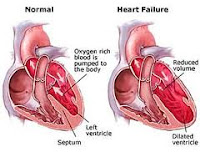
Illustrations
Thorax, normally
Heart failure, severe
Heart disease long term effects

Facts
  • Condition in which the heart fails to pump blood around the body strong enough.
  • The most typical problems that arise, shortness of breath, decreased physical performance, lethargy, increased fatigue and decreased appetite.
  • Eventually, it may develop swelling in the legs and elsewhere in the body.
  • Different types of heart disease can cause heart failure, most commonly, post-myocardial infarction.
  • Years of high blood pressure can also be hard on the heart and cause heart failure.
  • Diabetes, infection, and alcohol can also weaken the heart.
  • Self-treatment combined with treatment in general practice or hospital.
  • The aim of treatment is to relieve symptoms, improve quality of life and increase life expectancy.

Disease condition of heart muscle

Disease condition of heart muscle


How is the condition?
Lighter grades of heart failure are treated in general practice. The pronounced heart failures are needed hospital treatment. The aim of treatment is to relieve symptoms, improve quality of life and increase life expectancy.

Self-Treatment
There are several things you can do:
Disease condition of heart muscle
  • Eat less salt. When you have a lower blood pressure, and make work easier for the heart. Salt bound fluid in the body, which increases the load on the heart. The total salt intake during the day should not exceed one teaspoon, corresponding to approx. 6 g of salt (NaCl). Limited intake of salt means cessation of all the extra salt added to the diet as in baking bread, potato cooking, etc.
  • Avoid large meals, eat less and do more frequent meals.
  • Avoid large amounts of alcohol.
  • Stump the smoke.
  • It may be helpful to lose weight if you are overweight.
  • Physical activity should be guided by symptoms. Can you regularly exercise a little; it is useful? However, you should not charge more than your body allows.
Pharmacotherapy
There are many drugs that are effective in heart failure. The major drug groups are:
  • ACE inhibitors work by widening blood vessels so that blood pressure goes down, and the load on the heart is reduced. This type of drug is today due to treatment in the vast majority of heart failure.
  • Beta blockers have the heart to beat slower, they lower blood pressure and strengthens the heart. Furthermore, this type of drug used by most people.
  • Diuretic reduces fluid in the body and thus the strain on the heart. It is part preferably in a form of triple therapy with ACE inhibitor and beta blocker.
  • Digitalis strengthens the heart. These were widely used before, but used less these days.
  • There are also other products that are beneficial in heart failure, and a host of new treatments is being developed and tested.
Blood thinning medications are used in some cases, especially if you have atrial fibrillation. Rhythm Stabilizing drugs are also used if you have the tendency to an irregular heartbeat.

Another treatment
For some it may be appropriate with surgery. This is particularly the flap failure. Among patients with a tendency to serious arrhythmias may be necessary to operate a pacemaker or electric equipment (ICD). Very rarely do a heart transplant.

How to diagnose heart failure

How the condition diagnosed?


The diagnosis will usually be suspected based on the typical symptoms are present. These are mentioned above. It will have blood tests to identify disease, and radiography of the chest is also relevant to distinguish heart disease from lung disease with similar symptoms. ECG may show signs of heart disease.

How to diagnose heart failure

Is there any doubt about the diagnosis, an ultrasound of the heart ( echocardiography ) to decide with certainty, whether there is heart failure or not? Occasionally, it may be necessary to do another research to clarify the cause of heart failure and the extent of the damage to the heart.

Causes Of Heart Failure

Causes Of Heart Failure


What causes heart failure?
When we are in motion, increases metabolism throughout the body. The different body parts need then more blood to get enough oxygen and nourishment. A healthy heart can increase their activity so that blood circulation is satisfactory. A diseased heart is not capable of this, and the condition known as congestive heart failure.

Different types of heart disease can cause heart failure, but the most common is that it occurs after myocardial infarction. A portion of the heart muscle is dead then, and it is replaced by a rigid scar tissue. This reduces the hearts pumping ability. Years of high blood pressure can also be hard on the heart and cause heart failure.
Causes Of Heart Failure

For the same reason, the failure of the heart valves ( aortic stenosis, aortic insufficiency, mitral stenosis, mitral regurgitation ) provide heart failure due to increased wear and tear of the muscle. There are also a number of conditions that weaken the heart (cardiomyopathy) such as diabetes, infection, alcohol.

Define Heart Failure

Define Heart Failure


What is heart failure?

Heart failure describes a condition where the heart fails to pump blood around the body strong enough. This means that each agency receives insufficient blood supply; the most typical problems that arise, the heavy breathing and impaired physical performance. For a mild degree of heart failure is marked by this first effort, while in severe heart failure have shortness of breath even at rest. You will also notice that breathing becomes heavier when you lie down, and it will be easier in an upright position. Eventually, it may develop swelling in the legs and elsewhere in the body. Other symptoms include weakness, increased fatigability, decreased appetite.
Define Heart Failure
Approximately, 50000-100000 have heart failure. Estimate that 10% over 75 years, congestive heart failure.

What is the prognosis

What is the prognosis


What is the prognosis?
Most case of acute pericarditis is a self-limiting, short-term course - often less than a week, and the prognosis is good. 10-30% of patients experience a relapse during the first months, but later decrease recurrence risk. Get (approx. 1%) developed constrictive pericarditis.
What is the prognosis
Bacterial pericarditis, which these days are a rare condition, may sometimes be life threatening. Furthermore, constrictive pericarditis can be life-threatening condition.

Treatment Pericarditis

Treatment Pericarditis


What is the treatment?
Patients with acute pericarditis usually observed at the hospital with ECG monitoring because it can be associated inflammation of the heart muscle (myocarditis) and thus the risk of an irregular heartbeat. Treatment is primarily symptomatic relief. Pain and fever treated with anti-inflammatory drugs (e.g. ibuprofen) and in most cases, colchicine tablets. Treatment usually continues for at least a week after symptoms are gone. Only exceptionally is cortisone treatment is required.
Treatment Pericarditis

If it accumulates a lot of fluid in pericardial, the patient must be monitored closely and evaluated for pericardial tapping, i.e., withdrawal of fluid from the cavity of the pericardium. Other rarer forms of pericarditis treated in relation to what is the underlying cause.

Symptoms Of Pericarditis

What are the symptoms of pericarditis?
Acute nonspecific pericarditis can occur without warning, or it can be assumed passed by a respiratory infection. Chest pain is the most frequent symptom and often occurs quite suddenly. The pain is usually intense, sharp and is located centrally or on the left chest. The pain sometimes radiates to the shoulders, neck, back, and possibly to the left arm, and can resemble the pain of acute myocardial infarction.

Typically, pericarditis merten is that it often gets worse when breathing deeply, coughing, swallowing or lying on your back. The pain is often relieved when sitting bent forward. The pain can last from quite a few hours to several days. Fever is often present, and there may be coughing and breathing difficulty.
Symptoms Of Pericarditis

In constrictive pericarditis, there is often a history of previous pericarditis, cardiac surgery or radiotherapy. Typical of this condition is increasing breathing difficulty, fatigue and weakness. The patient can tolerate little physical activity and are quickly out of breath with exertion.

What Causes Pericarditis

What Causes Pericarditis


What are the causes?
The main causes of acute pericarditis are viral infections (> 80%). Pericarditis can also be seen in tuberculosis or HIV infection (common in developing countries), or in connection with cancer. Other possible causes include kidney failure, and radiation damage.

In 5-10% of patients with acute myocardial infarction occurs a slight pericarditis after a few days. The condition is usually harmless, and it goes back by itself. As a result of modern treatment (thrombolysis), this pericarditis form becomes less frequent. Another type of pericarditis occurs later in throughout heart attack in 10-20% of infarct patients.
What Causes Pericarditis
The chronic constrictive form can occur as a result of radiotherapy (radiation to the tumor tissue in the chest cavity), heart surgery or previously performed viral pericarditis.

Inflammation Of Heart

Inflammation Of Heart


Pose Heart inflammation - pericarditis

What is pericarditis?
The heart is surrounded by a thin membrane called the pericardium or pericardium. This membrane consists of two layers an inner membrane that attaches to the surface of the heart muscle (myocardium), and an outer membrane. In between the two membrane is a tiny amount of fluid that allows the two membranes to glide smoothly against each other when the heart is working.

A pericarditis is an inflammation of the pericardium. The distinction between three main forms of pericarditis:

An acute nonspecific pericarditis which manifests itself as a mild inflammation around the heart sac, and probably in most cases caused by a viral infection.
Inflammation Of Heart

Special and rare forms of acute pericarditis that may be caused by bacterial infection, immune disorder, or that may occur after a heart attack.

Chronic constrictive pericarditis is rare sequelae of acute pericarditis in which healing has caused scarring, and the pericardium has become stiff and tight. This may hamper the heart's motion and thereby impair the heart's ability to pump blood.

A pericarditis is a relatively rare condition. The acute non-specific form is the most frequent variant and accounts for more than 80% of all cases. It is seen at all ages but is most common in 15-30 years of age and most commonly in men.

Myocarditis Diagnosis

Myocarditis Diagnosis


How is the diagnosis?
Medical history suggestive of the condition. At the medical examination, it is classic heart failure signs bulging veins on his neck and crackles over the lungs.

Damage to heart muscle cells gives rise to certain blood tests (cardiac enzyme such as CK-MB and troponin-I) which is also elevated in myocardial infarction. The difference is that the heart muscle inflammation keeps these blood samples is increased further. Viral and bacterial tests can confirm that the patient has recently had or have a viral or bacterial infection. ECG may show transient light rhythm with extra strokes. Life threatening arrhythmias may occur late or early in the course.
Myocarditis Diagnosis

Radiographs of the chest may show diffuse enlargement of the heart shadow and fluid in the lungs. Echocardiography will immediately determine the severity and can judge the hearts pumping function. Other imaging modalities are rarely used in suspected acute myocarditis, but MRI is shown to be beneficial in some cases.

Diagnosis can be confirmed in difficult cases by taking a tissue sample (biopsy) of heart muscle tissue. The disadvantage is that inflammatory changes may be patchy in the heart, and it is not certain that the sample is taken from the inflamed tissue. Such sampling is not entirely harmless.

Myocarditis Signs And Symptoms

Myocarditis Signs And Symptoms


Symptoms and signs

The diagnosis is in many cases difficult to ask. Disease signs may vary from enlarged lymph nodes and non-specific flu-like symptoms of acute heart failure with great difficulty breathing or sudden death. In many cases, the disease is so little said that the precursor without symptoms. The symptoms can appear several days to weeks after the start of acute febrile illness or respiratory infection.
Myocarditis Signs And Symptoms

Typical symptoms include rapid heartbeat, and possibly chest pain. Other signs include unexplained heart failure and heart rhythm disorders that may cause fainting. Myocarditis can cause chest pain and, therefore, similar to acute myocardial infarction. Details of previous respiratory distress, gastrointestinal symptoms or febrile illness may help to estimate the time from viral infection of heart muscle inflammation appears.

Acute myocarditis should be suspected in young, especially males, who have had heart disease without certain evidence, such as heart failure, myocardial infarction, cardiac arrhythmias or conduction disturbances.

Myocarditis Causes

Myocarditis Causes


What causes myocarditis?
It is, in most cases, talk of an immune response during an infection, that is an indirect ( autoimmune ) response to infection. Only in rare cases, it involves the direct spread of viruses and bacteria that cause inflammation of the heart muscle (infection).

In our part of the world occurs heart muscle inflammation usually assuming viral infection in developing countries, both viruses, bacteria, parasites and rheumatic fever cause.
Myocarditis Causes

Inflammation of the heart can cause a clinical picture ranging from acute heart failure to barely noticeable disease - and fortunately, the cases with little disease the most frequent. Apart from serious pump failure, the disease also led to severe arrhythmia of the heart. Acute myocarditis is often completely over, but it may pass into an incurable phase even after the causative agents have disappeared. Chronic myocarditis can lead to enlarged and flabby heart (dilated cardiomyopathy), where the patient develops increasing symptoms of heart failure over several years.

What is myocarditis

What is myocarditis?


Myocarditis (myocarditis) is defined as an inflammation of the heart muscle. The inflammation may be caused by viruses or bacteria, toxins (toxins) or autoimmune reactions - that is, the body develops antibodies that attack the heart muscle. The condition leads to impaired function of the heart.
What is myocarditis

Myocarditis is not a common disease, but it is probably not so rare either. Accurate informatinn on the presence missing because the disease often completed without the disease is detected and goes by itself. Myocarditis is one of the most frequent causes of sudden death in young apparently healthy people.

What Is Chest Wall Pain

What Is Chest Wall Pain?


Chest Wall Pain
  • Chest wall pain caused by pain in the muscles between the ribs of the chest wall.
  • It is a common condition that often occurs in adulthood, with no significant gender difference.
  • Typical complaints of patients stinging pain, there is local muscle tension, and it is tender to touch the chest wall. The patient does not have a cough.
  • The patient needs no treatment other than information about the condition is harmless, stress-related and will be able to heal by itself.
  • For many this is a condition that comes and goes.
What Is Chest Wall Pain
How can the condition be treated?
This is a harmless condition that needs treatment. The important thing is to rule out other diseases and become familiar with the fact that there is nothing wrong with the heart or lungs. Most people will also benefit from relaxation exercises and learning good breathing technique.

Very few patients can get more serious and long-term ailments. In such cases, treatment (transverse friction) physiotherapy help. If you constantly experience new periods of pain, it is important to be aware of the cause and the good prognosis. Concerns increase muscle tension and therefore, pain.

What is the prognosis?
In the aforementioned German / Austrian study found that over half of the patients still had chest wall pain six months after the diagnosis was made. Complaints will often come and go, and in many cases. There is clear correlation between symptoms and stress-filled life phases.